Article Text

Abstract
Introduction The speed of declining kidney function differs among patients with diabetic nephropathy. This study was undertaken to clarify clinical and pathological features that affect the speed of declining kidney function in patients with diabetic nephropathy.
Research design and methods This study was design as multicenter retrospective study. The subjects (377 patients with diabetic nephropathy diagnosed by kidney biopsy at 13 centers in Japan) were classified into three groups based on the estimated glomerular filtration rate (eGFR) declining speed. The eGFR increasing group, the control group, and the eGFR declining group were divided at 0 and 5 mL/min/1.73 m2/year, respectively. Characteristics of clinicopathological findings of declining kidney function were evaluated.
Results The mean observation period of this study was 6.9 years. The control group, the eGFR increasing group, and the eGFR declining group included 81, 66, and 230 patients, respectively. The incidences of composite kidney events represented by 100 persons/year were 25.8 in the eGFR declining group and 2.0 in the eGFR increasing group. After adjustment for age, sex, systolic blood pressure, hemoglobin, and urinary albumin levels, three clinicopathological findings (urinary albumin levels, presence of nodular lesion, and mesangiolysis) were risk factors for inclusion in the eGFR declining group (the ORs were 1.49, 2.18, and 2.08, respectively). In contrast, the presence of subendothelial space widening and polar vasculosis were characteristic findings for inclusion in the eGFR increasing group (the ORs were 0.53 and 0.41, respectively).
Conclusions As well as urinary albumin elevation, nodular lesion and mesangiolysis were characteristic pathological features of patients with fast declining kidney function.
- nephropathology
- nephropathy
- kidney biopsies
- kidney failure
This is an open access article distributed in accordance with the Creative Commons Attribution Non Commercial (CC BY-NC 4.0) license, which permits others to distribute, remix, adapt, build upon this work non-commercially, and license their derivative works on different terms, provided the original work is properly cited, appropriate credit is given, any changes made indicated, and the use is non-commercial. See: http://creativecommons.org/licenses/by-nc/4.0/.
Statistics from Altmetric.com
Significance of this study
What is already known about this subject?
Some part of diabetic cases exhibit rapid reduction in kidney function and reach end-stage renal disease (ESRD) in a short period of time, and they are called “rapid eGFR decliner”.
Although many clinical studies have been performed regarding kidney prognosis in patients with diabetic nephropathy, the pathological features of rapid decliners have not been fully elucidated.
What are the new findings?
This study revealed that as well as urinary albumin elevation, two pathological findings—nodular lesion and mesangiolysis—were characteristic features of patients with rapidly declining kidney function.
Two pathological findings—subendothelial space widening (or duplication of basement membrane) and polar vasculosis—were characteristic features of patients who could recover their kidney function after kidney biopsy.
The incidences of composite kidney events (new-onset ESRD, reduction of eGFR by ≥50%, or doubling of the serum creatinine level) in the eGFR declining group were more than 10 times higher than that in the eGFR increasing group.
How might these results change the focus of research or clinical practice?
Among the various pathological changes in diabetic kidney disease (DKD), this study clarifies the important pathogenic prognosis features in rapid eGFR decliner, and these features should be therapeutic target for DKD.
This study also will give a clue to detect good biomarkers associated with characteristic pathogenic change in rapid eGFR decliner.
Introduction
The speed of declining kidney function differs among patients with diabetic nephropathy.1–3 There are two characteristic groups: “rapid decliners” and “no or slow decliners”.4–6 In addition, some patients recover kidney function.4 Fast decliners exhibit fast reduction in kidney function and reach end-stage renal disease (ESRD) in a short period of time, whereas slow decliners preserve a degree of kidney function for an extended period of time. Advanced proteinuria is a key clinical factor in the detection of fast decliners.1 Although the reduction of kidney function is usually followed by massive proteinuria, some patients show progressive kidney dysfunction with low-grade proteinuria. Therefore, further factors in addition to proteinuria are required. Although many clinical studies have been performed regarding kidney prognosis and cardiovascular events in patients with diabetic nephropathy,7 8 the pathological features of fast decliners have not been fully elucidated.
In recent years, many patients have been diagnosed with diabetic kidney disease without kidney biopsy assessment. However, pathological evaluation should be essential to understand and recognize the specific disease condition and activity of each patient. In many countries, the accumulation of evidence for diabetes treatment and the use of new drugs have enabled patients with diabetes to achieve glycemic control.9–11 Moreover, various medication, including renin/angiotensin system inhibitors, modified the levels of proteinuria and clinical manifestation. Furthermore, hypertension, dyslipidemia, aging, and other factors modify pathological progression in diabetic kidney disease.12 These various conditions would make it difficult to speculate the disease progression status. Therefore, pathological assessment in addition to clinical parameters, including proteinuria, should be required to understand the disease condition and activity of each patient. We previously reported differences and similarities between patients with diabetic nephropathy and patients with hypertensive nephrosclerosis.13 14 Moreover, we have reported the importance of histological analysis in addition to clinical stage to predict the kidney prognosis or cardiovascular events in patients with diabetic nephropathy.15–17 From this perspective, pathological evaluation of diabetic patients is of considerable value.
Because many patients are diagnosed with diabetic nephropathy, it is impossible to provide uniform and intensive care of all affected patients. Therefore, there is a need to identify patients at high risk of progressive kidney dysfunction and to closely monitor these patients. In the present study, we aimed to clarify the clinical and pathological features of patients with fast declining kidney function, as well as patients who exhibited recovery of kidney function. This analysis revealed that urinary albumin elevation and two pathological findings—nodular lesion and mesangiolysis—were characteristic features of patients with fast declining kidney function. Moreover, two pathological findings—subendothelial space widening (or duplication of basement membrane) and polar vasculosis—were characteristic features of patients who exhibited recovery of kidney function.
Methods
Study design and study population
This study was design as multicenter retrospective study. The subjects were 600 patients who had been diagnosed with diabetic nephropathy by kidney biopsy during the period from January 1, 1985 to December 31, 2016; samples were collected by our study group of the Ministry of Health, Labour and Welfare, as well as the Agency for Medical Research and Development in Japan. Data regarding 377 patients for whom the reduction of kidney function could be calculated using follow-up data for 3 years after kidney biopsy, and who had estimated glomerular filtration rate (eGFR) >15 mL/min/1.73 m2, were analyzed in this study. The indications for biopsy were kidney function impairment and urinary abnormalities, such as albuminuria, proteinuria, hematuria, or abnormal casts in urine. Patients were excluded if they had a diagnosis of concomitant kidney disease with diabetic kidney disease and/or if they underwent protocol kidney transplant biopsy. Type 2 diabetes was defined as having diabetes onset after age 30 years and not taking insulin at the initial visit in our hospitals.
The patients were classified into three groups based on the rate of decline in kidney function. In accordance with KDIGO guidelines’ definition of progression, a rate of eGFR decline exceeding 5 mL/min/year was used as a cut-off.18 Thus, the control group was defined as ≥0 and <5 mL/min/1.73 m2/year in mean eGFR decline within 3 years after kidney biopsy; the eGFR increasing within 3 years after biopsy group (eGFR increasing group) was defined as <0 mL/min/1.73 m2/year in mean eGFR decline within 3 years after kidney biopsy; and the eGFR declining within 3 years after biopsy group (eGFR declining group) was defined as ≥5 mL/min/1.73 m2/year in mean eGFR decline within 3 years after kidney biopsy. The control group included 81 patients, the eGFR increasing group included 66 patients and the eGFR declining group included 230 patients. We compared the control group with the eGFR increasing and eGFR declining groups. Clinical data were used at the time of kidney biopsy and clinical follow-up. Histological evaluations were performed in accordance with the method used in a previous paper.16 Clinically required biopsy samples were used. Under the approval of the ethics committee of Kanazawa University, the opt-out approach is used in this study. Moreover, this study was conducted in accordance with Declaration of Helsinki.
Pathological examinations
Biopsy samples were stained with periodic acid–Schiff reagent, periodic acid–methenamine silver, H&E, and Mallory-Azan or Masson’s Trichrome stains, then examined by light microscopy. In accordance with a previous study, nine glomerular lesions, two interstitial lesions, and two vascular lesions were evaluated in each biopsy sample.16 The scoring system is as follows: nine glomerular lesions (diffuse lesion (grades 0–3); nodular lesion (grades 0–1); subendothelial space widening (grades 0–3); exudative lesion (grades 0–1); mesangiolysis/microaneurysm (grades 0–1); perihilar neovascularization (grades 0–1); global glomerulosclerosis, collapsing glomerulopathy, and ischemic nephropathy (grades 0–1); segmental sclerosis (grades 0–1); glomerulomegaly (grades 0–1)), two interstitial lesions (interstitial fibrosis and tubular atrophy (grades 0–3) and interstitial cell infiltration (grades 0–3)), and two vascular lesions (arteriolar hyalinosis (grades 0–3) and intimal thickening (grades 0–3)). We also evaluated the percent glomerular sclerosis as defined by the number of total global and segmental sclerotic glomeruli per total number of glomeruli. Pathologists in each center performed all pathological scoring in this study.
Clinical data
Age, sex, body mass index, systolic blood pressure, diastolic blood pressure, hemoglobin (Hb) A1c, and total cholesterol were used as baseline clinical parameters at the time of the kidney biopsy. eGFR was calculated using the following formula: eGFR (mL/min/1.73 m2)=194×serum creatinine−1.094×age−0.287.
(For female patients, this value was multiplied by 0.739.)
HbA1c levels were recorded as national glycohemoglobin standardization program values, in accordance with the recommendations of the Japanese Diabetic Society and the International Federation of Clinical Chemistry.
Outcomes
The primary outcome of this study was composite kidney events, which were defined as new-onset ESRD, reduction of eGFR by ≥50%, or doubling of the serum creatinine level. ESRD was defined as initiation of hemodialysis/peritoneal dialysis, renal transplantation, or death as a result of uremia. The secondary outcomes for this study were kidney death (hemodialysis/peritoneal dialysis, renal transplantation, or death as a result of uremia), cardiovascular events (cardiovascular death, nonfatal myocardial infarction, coronary intervention, or nonfatal stroke), and all-cause mortality. The sources of the outcome data were collected from the medical records of each center. Patients who did not reach the outcome of interest or who were lost to follow-up were censored at their last follow-up visit.
Statistical analysis
Data are expressed as mean±SD or median (IQR). Continuous variables were compared between groups using the Mann-Whitney U test for nonparametric data; categorical variables were compared between groups using the χ2 test. To minimize the clinical background difference between groups, propensity score method was used. Patients’ background characteristics, including urinary albumin, Hb, and systolic blood pressure, were balanced in the analysis. Cox regression analysis was used to calculate adjusted HRs for each outcome. Survival curves were obtained using Kaplan-Meier analysis and compared using the log-rank test. Univariate and multivariate logistic regression models were used to analyze factors for eGFR declining and eGFR increasing groups. All analyses were conducted using Stata V.13 (StataCorp, College Station, TX, USA). A two-sided p value of <0.05 was considered statistically significant.
Results
Background of clinical manifestations in each group
The eGFR increasing, control, and eGFR declining groups included 66, 81, and 230 patients, respectively (online supplementary figure 1). The mean observation period of this study was 6.9 years. The median baseline eGFR in the eGFR increasing group was 53.9 mL/min/1.73 m2, whereas it was 50.2 mL/min/1.73 m2 in the control group; conversely, the median baseline eGFR in the eGFR declining group was 47.3 mL/min/1.73 m2. Table 1 summarizes the clinical manifestations of the three groups. When comparing the eGFR increasing and eGFR declining groups with the control group, there were no differences in proportion of men, body mass index, diastolic blood pressure, eGFR, HbA1c level, or hematuria-positive rate. However, systolic blood pressure and urinary albumin levels increased among groups: eGFR increasing (lowest), control, and eGFR declining (highest). Serum levels of total protein and albumin decreased in the eGFR declining group, compared with the control group, as did the Hb level; serum levels of total cholesterol increased in the eGFR declining group, compared with the control group. Regarding medication usage, renin/angiotensin system inhibitors were less frequently used in the eGFR increasing group. Systolic and diastolic blood pressures, as well as serum total protein, serum albumin, and urinary protein levels were strongly correlated (online supplementary table 1). In terms of clinical classification, approximately 60% of patients were classified into chronic kidney disease Category Red, and approximately 80% of patients were stage 3 of diabetic nephropathy based on the Japanese classification 2014 (online supplementary table 2, online supplementary figure 2).19
Supplemental material
Baseline clinical characteristics stratified by eGFR declining speed
Clinical outcomes in each group
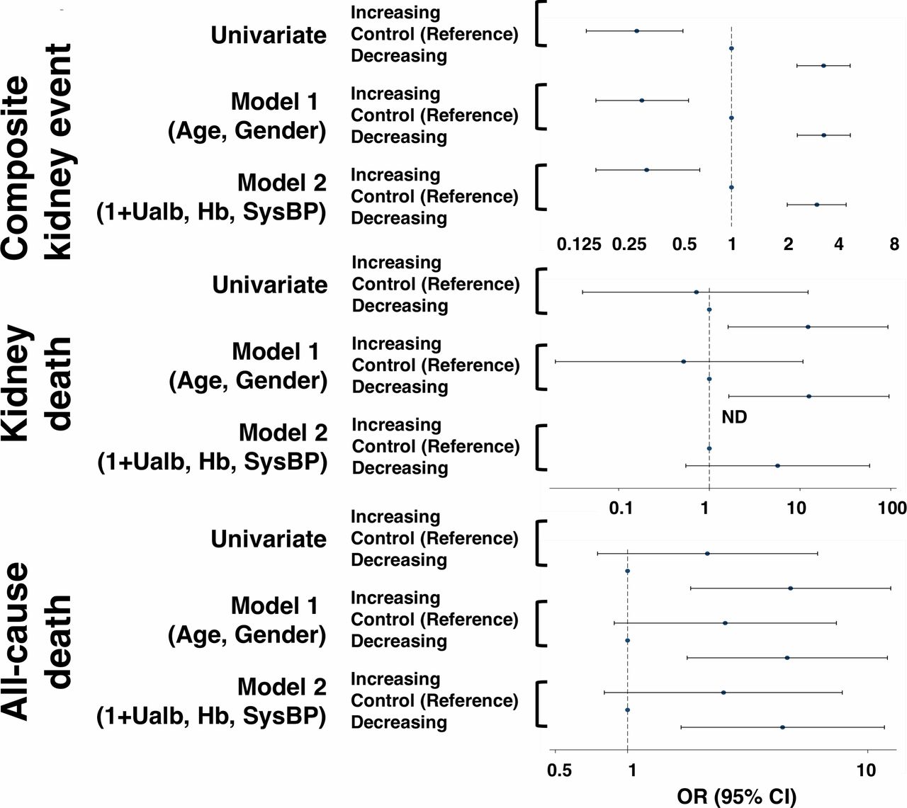
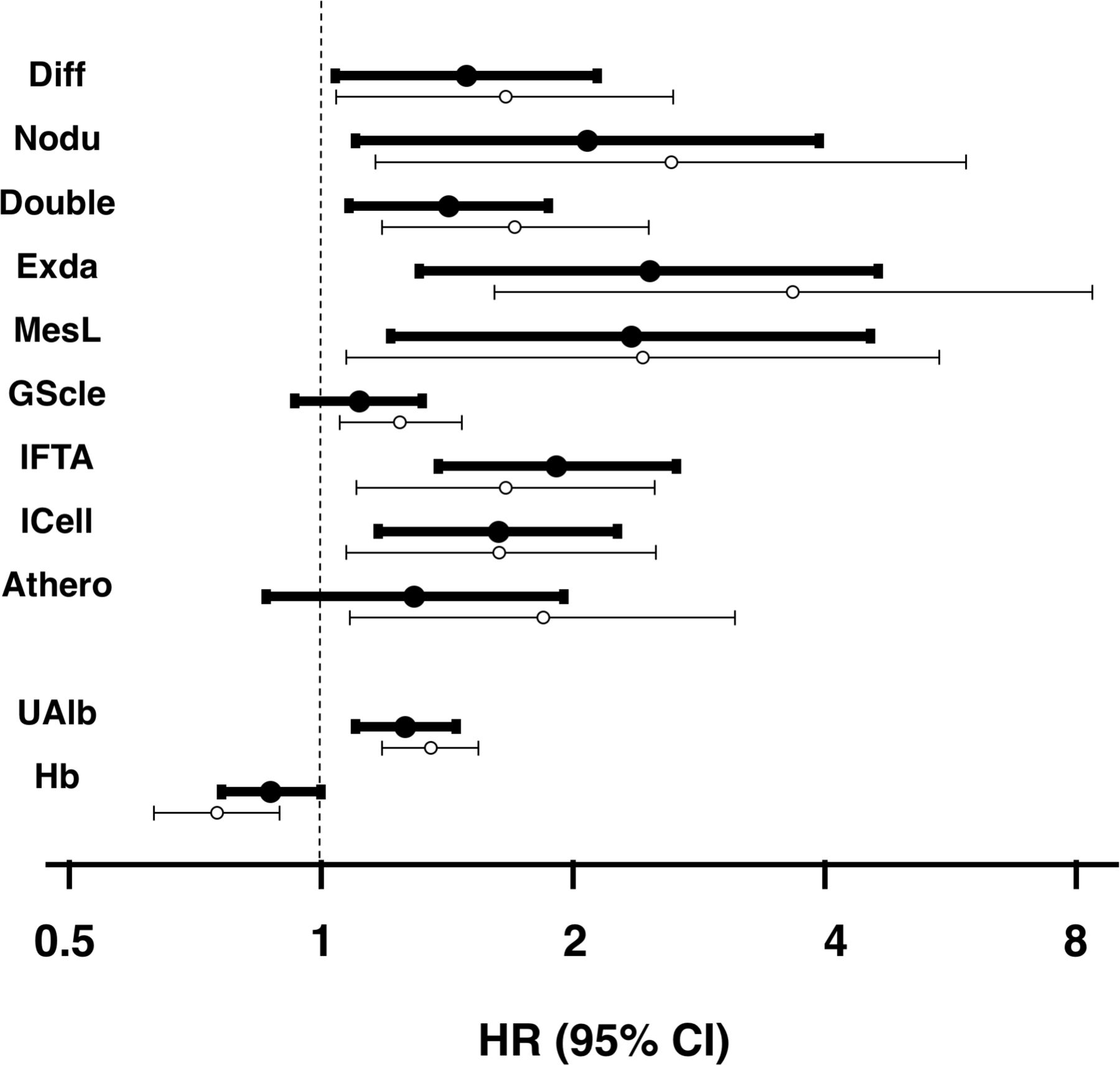
The incidences of composite kidney events and kidney deaths, represented by 100 persons/year, were 25.8 and 3.0 in the eGFR declining group, respectively. In contrast, the incidences of those outcomes were 2.0 and 0.2 in the eGFR increasing group (online supplementary table 3). Both incidence rates were approximately 10 times higher in the eGFR declining group than in the eGFR increasing group. Moreover, respective rates of death and cardiovascular events were 2.9 and 3.3 in the eGFR declining group, whereas they were 1.7 and 1.1 in the eGFR increasing group. After adjustment for age, sex, urinary albumin level, Hb and systolic blood pressure, the adjusted HRs for composite kidney events in Cox analysis were 3.05 and 0.31 in the eGFR declining and eGFR increasing groups, respectively (figure 1). The adjusted HR for all-cause mortality was also high in the eGFR declining group (figure 1). The survival curve of composite kidney events in each of the three groups are shown by the Kaplan-Meier curve (figure 2). In the eGFR declining group, approximately 50% of patients had composite kidney events within 3 years and 80% had the events within 8 years after kidney biopsy. Conversely, in the eGFR increasing group, >50% of patients had no composite kidney events within 20 years after kidney biopsy. Kidney death showed a similar pattern to that of composite kidney events (online supplementary figure 3). In addition to the Kaplan-Meier analysis, crude and adjusted HR for kidney death were evaluated by the Cox analysis (figure 3). Seven glomerular and interstitial pathological changes, including exudative lesion, mesangiolysis, nodular lesion, and interstitial fibrosis, and two clinical data, including urinary albumin levels and anemia levels, were detected as risk factors for kidney death in multivariable analysis adjusted for age, gender, urinary levels of albumin, and hemoglobin.

Crude and adjusted HR for composite kidney event, kidney death, and all-cause death, among three groups by eGFR declining speed. Cox analysis indicated that eGFR increasing group and eGFR declining group were statistically significantly high and low risk for composite kidney event even after adjusted by age, gender, urinary albuminuria, Hb, and systolic pressure, respectively (control group is reference). Similarly, eGFR declining group was statistically significantly high risk for all-cause death even after adjusted by age, gender, urinary albuminuria, Hb, and systolic pressure. eGFR, estimated glomerular filtration rate; Hb, hemoglobin; ND, not determined; SysBP, systolic blood pressure; Ualb, urinary levels of albumin.

Survival curves for composite kidney end points stratified by three groups as derived from the Kaplan-Meier method with log-rank test. Event-free curves of composite kidney end points are shown. Differences between two groups were compared by a log-rank test.

Crude and adjusted HR for kidney death. Crude and adjusted HR for kidney death were determined for various pathological clinical characteristics (indicated in the figure) by the Cox analysis. A plot of the HR for each variable is shown; open circles indicate the HR in univariate analysis, and solid circles indicate the HR in multivariable analysis with age, gender, urinary levels of albumin, and hemoglobin; 95% CI shown as whiskers. HRs were calculated for a one score increase in each pathological finding; 10% increase in global glomerulosclerosis; 1 g/day increase in urinary levels of albumin; 1 g/dL increase in Hb. Athero, intimal thickening; Diff, diffuse lesion; Double, subendothelial space widening (or duplication of basement membrane); Exda, exudative lesion; GScle, global glomerulosclerosis, collapsing glomerulopathy, and ischemic nephropathy; Hb, hemoglobin; ICell, interstitial cell infiltration; IFTA, interstitial fibrosis and tubular atrophy; MesL mesangiolysis/microaneurysm; Nodu, nodular lesion; UAlb, urinary levels of albumin.
Pathological changes in each group
The incidence of each pathological finding increased as follows: eGFR increasing group (lowest), control group, and eGFR declining group (highest) (table 2, online supplementary figure 4). However, after adjustment using a propensity score matching system with Hb, systolic blood pressure, and urinary levels of albumin, there were no statistical differences between the control and eGFR declining groups (table 2). Three pathological findings were significantly different between the eGFR increasing group and control groups even after matching: subendothelial space widening (or duplication of basement membrane), global sclerosis, and interstitial cell infiltration. These findings were confirmed when our cohort was divided into two group by ≥5 mL/min/1.73 m2/year in mean eGFR decline within 3 years after kidney biopsy (online supplementary table 4). After adjustment using a propensity score matching system with Hb, systolic blood pressure, and urinary levels of albumin, there were no statistical differences between the two groups (online supplementary table 4B).
Baseline pathological characteristics stratified by eGFR declining speed
Predictive factors for inclusion in the eGFR declining group
In contrast to the baseline findings, these pathological findings influenced whether patients were in the eGFR declining or eGFR increasing groups. Using multivariate logistic analysis, risk factors for eGFR declining and eGFR increasing groups were evaluated, in comparison with control. In univariate analysis, various clinicopathological findings increased the risk of inclusion in the eGFR declining group, and reduced the risk of inclusion in the eGFR increasing group, relative to the control group (figure 4A). After adjustment for age, sex, systolic blood pressure, Hb levels, and urinary albumin levels, three clinicopathological findings (urinary albumin levels, presence of nodular lesion, and presence of mesangiolysis) were risk factors for inclusion in the eGFR declining group; the ORs were 1.49, 2.18, and 2.08, respectively. In contrast, the presence of subendothelial space widening (or duplication of basement membrane) and presence of polar vasculosis reduced the risk for inclusion in the eGFR increasing group, relative to the control group; the ORs were 0.53 and 0.41, respectively (figure 4B). When examining these four factors using receiver operating characteristic curve analysis, the areas under the curve of urinary level of albumin, the presence of nodular lesion, and the presence of mesangiolysis were 0.75, 0.62, and 0.61, respectively. Moreover, the areas under the curve of the presence of subendothelial space widening (or duplication of basement membrane) and the presence of polar vasculosis were 0.63 and 0.66, respectively.

OR to the risk for inclusion in the eGFR decreasing group compared with control group (A) and OR to the risk for inclusion in the eGFR increasing group compared with control group (B). ORs to the risk for inclusion in the eGFR decline group or eGFR increasing group were determined for various pathological and clinical characteristics (indicated in the figure) by the logistic model. A plot of the OR for each variable is shown; open circles indicate the OR in univariate logistic regression, and solid circles indicate the OR in multivariable logistic regression; 95% CI shown as whiskers. ORs were calculated for a one score increase in each pathological finding; 10% increase in global glomerulosclerosis; 1 g/day increase in urinary levels of albumin; 1 g/dL increase in Hb; 10 mm Hg increase in systolic blood pressure. Diff, diffuse lesion; Double, subendothelial space widening (or duplication of basement membrane); eGFR, estimated glomerular filtration rate; Exda, exudative lesion; GScle, global glomerulosclerosis, collapsing glomerulopathy, and ischemic nephropathy; Hb, hemoglobin; Hyali, arteriolar hyalinosis; ICell, interstitial cell infiltration; IFTA, interstitial fibrosis and tubular atrophy; MesL, mesangiolysis/microaneurysm; Nodu, nodular lesion; Pola, perihilar neovascularization (polar vasculosis); SysBP, systolic blood pressure; uAlb, urinary levels of albumin.
These findings were confirmed when our cohort was divided into two groups by ≥5 mL/min/1.73 m2/year in mean eGFR decline within 3 years after kidney biopsy (online supplementary figure 4). After adjustment for age, sex, systolic blood pressure, Hb levels, and urinary albumin levels, three clinicopathological findings (urinary albumin levels, presence of nodular lesion, and presence of mesangiolysis) were included in risk factors for inclusion in the eGFR declining group.
Discussion
In this study, we aimed to elucidate the clinicopathological factors that contribute to fast declining kidney function. The clinical features of the group with fast declining kidney function were high urinary albumin, hypertension, and anemia. Analysis of pathological factors revealed that the presences of nodular lesions and mesangiolysis were independently associated with fast declining kidney function. Moreover, the eGFR increasing group had reduced incidences of the presence of polar vasculosis and the presence of subendothelial space widening (or duplication of basement membrane). These pathological changes might contribute to the different types of pathogenesis that characterize the progression of diabetic kidney disease.
Nodular lesions and mesangiolysis were independent pathological risk factors for fast declining kidney function. In our previous studies, patients with nodular lesions or mesangiolysis exhibited poor kidney prognosis, despite the absence of albuminuria.14 15 Moreover, there was a strong positive correlation between the presence of nodular lesions and the presence of mesangiolysis.13 Type VI collagen is reportedly accumulated in nodular lesions; this type of collagen is resistant to various intrinsic collagenases.20 21 Moreover, mesangiolysis is a typical pathological change associated with reduction of the glomerular filtration surface in the glomerular capillary lumen.22 These pathological changes were expected to be predictive factors of declining kidney function and should be key targets for treatment of diabetic kidney disease.23 24 However, the mechanisms by which nodular lesions and mesangiolysis occur in patients with diabetic nephropathy are not yet clear. There are limited animal models of diabetic nephropathy with nodular lesions and mesangiolysis21 25; thus, additional research is urgently needed.
Polar vasculosis and subendothelial space widening (or duplication of basement membrane) have other clinical impacts on declining kidney function. Importantly, these pathological findings were able to distinguish the eGFR increasing group and control groups. There have been only a few reports regarding the clinical significance of polar vasculosis.16 26 27 Polar vasculosis is expected to connect glomerular and peritubular capillaries. However, the details of its structure and function remain unclear. Moreover, subendothelial space widening (or duplication of basement membrane) is a pathological change due to glomerular capillary endothelial cell dysfunction.28 The frequency of these two pathological changes gradually increases in accordance with the progression of albuminuria and reduction of eGFR. However, we identified patients without these pathological findings, who comprised the eGFR increasing group. It is unclear whether patients who recovered their kidney function after kidney biopsy were resistant to glomerular capillary endothelial cell dysfunction or neovasculosis, or whether these two pathological changes were predictive of declining kidney function. These points will be clarified by future investigations. Basic research, including animal experiments, and prospective clinical studies with therapeutic interventions are needed to elucidate the mechanisms and pathogenic implications of nodular lesions, mesangiolysis, polar vasculosis, and subendothelial space widening (or duplication of basement membrane) in diabetic kidney disease.
The prediction of declining kidney function without pathological findings is an important goal. In this study, patients with declining kidney function showed abundant urinary albumin, hypertension, and low Hb levels, compared with patients in the control group; urinary albumin elevation was also identified as an independent risk factor for declining kidney function. Furthermore, approximately 80% of patients in the eGFR declining group had stage 3 diabetic nephropathy based on the Japanese classification (albuminuria of >300 mg/g Cr and eGFR of >30 mL/min/1.73 m2). Many studies have shown that albumin is a predictive factor for kidney prognosis; our data are consistent with those previous findings.29–31 The clinical indicators identified in this study should be valuable for identification of patients with fast declining kidney function and can enable close monitoring to prevent disease progression in those selected patients. Moreover, to detect good biomarkers associated with pathogenesis of the characteristic pathological change in patients with fast decliners should be valuable in clinical setting of follow-up the patient with diabetic kidney disease.
This study had some limitations. It was retrospective and therefore included many biases. Selective bias due to kidney biopsy was presumably the most influential bias in this study. Moreover, we used reduction in eGFR ≥5 mL/min/1.73 m2/year as a cut-off criterion for inclusion of patients in the eGFR declining group in this study; other cut-off criteria could have been selected. We observed that a cut-off criterion of reduction in eGFR ≥10 mL/min/1.73 m2/year yielded results similar to those described in this paper (data not shown). In addition, the analysis of therapeutic points was limited because there was limited information available regarding treatment in this data set. Prospective research with treatment intervention is necessary, as are validation studies in different cohorts.
Comparison of the three groups based on mean eGFR decline revealed two characteristic pathological changes: the presences of nodular lesion and mesangiolysis were predictive of fast declining kidney function, whereas the presences of subendothelial space widening (or duplication of basement membrane) and polar vasculosis were predictive of kidney function recovery. These are valuable new findings that reveal the underlying pathogenesis of diabetic kidney disease. These pathological changes could be targets for future biomarker development and therapeutic interventions.
Acknowledgments
The authors thank Dr. Hiroshi Kida (National Hospital Organization Kanazawa Medical Center) for his helpful comments on this study.
References
Footnotes
Contributors The authors’ responsibilities were as follows: KF, YUe, SM, and HM conceived the study. YY, HK, YSu, NU, YUb, SN, TN, KS, KK, HY, and YSh participated in the collection and statistical analysis of the data. MY, NS, YI, TT, SK, and AH participated in the interpretation of the results. KF, MS, JH, and HS drafted the manuscript. KF and TW critically reviewed the manuscript. All authors read and approved the final manuscript. KF and TW are the guarantors of this work and, as such, had full access to all of the data in the study and take responsibility for the integrity of the data and the accuracy of the data analysis.
Funding This study was supported in part by a Grant-in-Aid for Diabetic Nephropathy and Nephrosclerosis Research from the Ministry of Health, Labour and Welfare of Japan and Grant-in-Aid for Practical Research Project for Renal Diseases, from the Japan Agency for Medical Research and Development (No. 15ek0310003h0001). This work was also supported in part by Grants-in-Aids from the Ministry of Education, Culture, Sports, Science, and Technology of the Japanese Government.
Competing interests None declared.
Patient consent for publication Not required.
Ethics approval Prior ethical approval was obtained from the Medical Ethics Committee of Kanazawa University (Approval No. 1204).
Provenance and peer review Not commissioned; externally peer reviewed.
Data availability statement All data relevant to the study are included in the article or uploaded as online supplementary information.